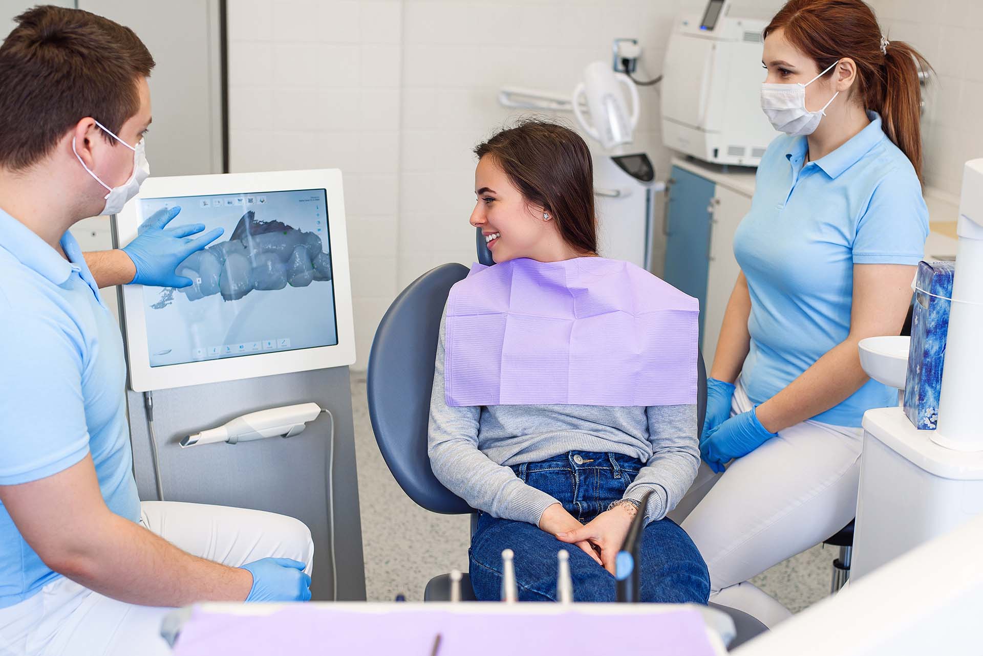
With the continuous development of computed tomography (CT) and the subsequent emergence of cone-beam computed tomography (CBCT), three-dimensional (3D) images can be provided more effectively, which has had a profound impact on surgical and medical practices. In oral and maxillofacial surgery, practitioners almost entirely relied on two-dimensional plain films in the past, while the applications and advantages of CBCT in dentistry are becoming increasingly prominent.
Meanwhile, 3D printing has achieved a considerable level of development in many industrial fields. In the medical field, 3D printing can be applied to diagnosis or treatment, and at the same time, tissues or organs can be 3D printed through cell bioprinting technology, which may change the paradigm of the medical field. This paper reviews the research progress of CBCT and 3D printing technology in maxillofacial surgery in recent years.
With the progress of basic scientific research in the field of CT-assisted oral and maxillofacial surgery, the functions of these technologies have been introduced into routine clinical practice. The 3D scanning segmentation technology for reconstructing soft and hard tissues is required for quickly switching between various modes and viewing superimposed images. CT, especially CBCT, is increasingly used in the study of facial and jawbone lesions.
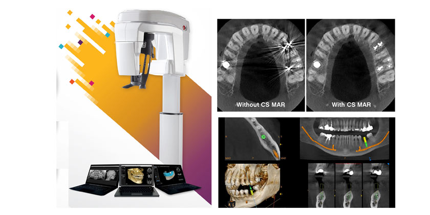
- Applications of CBCT in Maxillofacial Surgery
Traditional CT can usually reconstruct tomographic images of the maxillofacial region in a timely and relatively accurate manner and make a diagnosis. However, CT scanning has many layers, and it is difficult to avoid the drawback of missing small lesions due to respiratory movement during tomographic CT scanning. Given that CBCT has higher resolution, lower radiation dose, and lower cost in maxillofacial region imaging, it can easily replace traditional CT. 3D imaging of maxillofacial cysts and tumors can provide surgeons with important information needed for planning surgery. Through volume analysis, it can help predict the number and volume of planned implants required for reconstruction.
CBCT data can also be used to create stereoscopic models of the region of interest. With the assistance of complex craniofacial reconstruction, 3D printing technology has recently gained a reputation in the medical and surgical fields. Nowadays, maxillofacial surgery can benefit from additive manufacturing (AM) in various aspects and different clinical cases, obtaining comprehensive information about the patient’s maxillofacial region to achieve the expected treatment goals.
1.1 Temporomandibular Joint (TMJ) Diseases
The diagnosis and treatment planning of TMJ diseases are usually very challenging. Although MRI is still the “gold standard” for imaging the tissues inside the TMJ, usually only the bone tissues are evaluated based on conventional panoramic X-ray films. Panoramic X-ray photographs can provide a two-dimensional overall image of the joint, but they have low sensitivity in evaluating the changes of the condyle, poor reliability, and low accuracy in evaluating the temporal tissues of the joint. The images provided by current CBCT machines have shown that a complete X-ray evaluation of the bone tissues of the TMJ can be carried out, and the generated images have a high diagnostic quality. Compared with traditional CT, due to the significantly reduced radiation dose and cost, CBCT may soon become the preferred research tool for evaluating bone changes in the TMJ.
1.2 Cleft Lip and Palate Sequential Treatment
Since patients with cleft lip and palate are too young and there is a risk of radiation exposure, CT is not routinely used. The timing of alveolar cleft repair is usually determined based on panoramic and occlusal films. Compared with traditional X-ray images, CBCT should be able to better evaluate the age of teeth, the position of the arch segment, and the size of the cleft. Volume analysis is expected to provide better predictions regarding the defect morphology and the volume of graft materials required for repair.

1.3 Orthognathic Surgery
Clinicians have long been evaluating the usefulness of 3D imaging in orthodontic and orthognathic surgeries, mainly focusing on the correlation between soft tissue and hard tissue changes. With the early 3D imaging studies laying the foundation for the current cephalometric analysis and prediction, it is considered that 3D models are used for orthodontic and orthognathic analysis and surgical prediction before and after surgery, but experimental verification of the measured landmarks and their relationships is required. Although it is as useful as cephalometric analysis, its imaging accuracy is insufficient in deformities such as hemifacial microsomia syndrome, severe facial asymmetry, and occlusal inclination. 3D imaging of hard and soft tissues makes all data available, enabling more precise surgical and treatment plans to be implemented.
1.4 Impacted and Supernumerary Teeth
Using CBCT to locate and evaluate the affected canines and supernumerary teeth seems to make the surgical process more effective and less invasive, while increasing 3D imaging significantly improves the identification efficiency of potential complications of the affected teeth. In addition, on-site evaluation not only reduces the probability of trauma and shortens the time but also is more complete. Seeing the anatomical structures adjacent to the region of interest from three dimensions can reduce the morbidity during surgery and potential complications, thereby helping to obtain better results.
In conclusion, with the continuous reduction of the cost of CBCT technology, it is only a matter of time before CBCT enters ordinary oral and maxillofacial surgery. Its strong diagnostic ability and low radiation dose will also help this technology become mainstream.
- 3D Printing
3D printing is a manufacturing process in which objects are manufactured in a layered manner during the fusion or deposition of different materials (such as plastics, metal, ceramic, powder, liquid, and even living cells) to construct 3D structures. This process is also known as rapid prototyping (RP), solid freeform technology (SFF), or AM. 3D printing methods and their applications and benefits in contemporary oral and maxillofacial surgery have been demonstrated.

Methods for treatment planning and simulation using rapid prototype models of 3D printing have been established before oral cancer surgery or orthognathic surgery to ensure more precise and safer surgeries. In addition, in the field of dental implantation, surgical scaffolds can be manufactured using CT images. For example, 3D printing may be used in fracture surgery or reconstruction surgery, making it easier to produce customized and reconstructed steel plates and perform morphological reconstruction of bone defect areas. The applications of 3D printing technology in oral and maxillofacial surgery are as follows.
2.1 Surgical Planning
Since 3D printing can more effectively distinguish between traumatic and pathological defects, it has been proven to enhance the diagnosis and treatment of the maxillofacial region, and this function enables precise decision-making. In the case of pathological lesions, 3D printing can present the spatial relationship with surrounding components, and these important visualizations can minimize surgical complications. Through 3D printing, surgeons can improve the feasibility and precision of surgery through visualization programs, making oral and maxillofacial surgeries more precise, conforming to preoperative predictions. 3D printing can quickly generate models with acceptable precision and structural details to achieve better results and shorten the operation time.
2.2 Trauma Surgery
3D printers can help treat trauma patients with recent or delayed fractures and defects. Different fractures of the maxillofacial structure can benefit from 3D printing, and orbital fractures are the best targets of these methods. These patients can be treated by 3D-customized reconstruction of orbital wall defects with titanium meshes or thin plates. Before the start of surgery, the titanium mesh or plate is precisely fitted on the 3D-printed replica to help shorten the time of general anesthesia.
Complex orbital anatomy makes it difficult to reconstruct orbital defects, and postoperative eyelid ptosis or diplopia always occurs when the orbital wall is not correctly reconstructed. Surgeons can solve these complications by using 3D-printed titanium meshes of the contralateral orbital anatomy. Evaluating the application of implants customized by the 3D-printing system in reconstructing blowout fractures of the orbital floor, the average orbital volume (OV) of the affected side after surgery was significantly reduced, and there was no difference in the corrected orbital OV compared with the healthy side.
There have been studies using 3D models to treat three patients with medial orbital wall fractures. Using the 3D model as a template, bone grafts from the zygomatic bone were easily and precisely measured and collected, achieving perfect adaptation and shortening the operation time.
2.3 Orthognathic Surgery
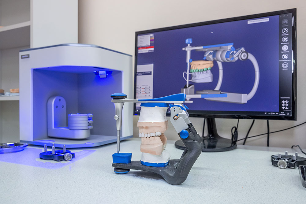
Precise planning and decision-making based on accurate diagnosis are crucial for the success of orthognathic surgery. As mentioned earlier, 3D printing technology shows some clinically obvious inaccuracies in orthognathic surgery, which is troublesome for ideal tooth occlusion.
2.4 Facial Prostheses
In the past decade, there have been reports of manufacturing prosthetic noses, ears, eyes, and faces. The literature indicates that better aesthetic and functional effects can be achieved through the application of 3D printing compared with traditional restoration. Facial prostheses manufactured by the RP method have been successfully utilized.
With the application of 3D printing technology, facial prosthetics has been widely developed. This technology can produce replicas of facial structures within just a few hours. AM technology is a common method for manufacturing facial prostheses. Long production times, soft tissue deformation, and patient discomfort are the main limitations of this process. Recently, 3D printing has been used to produce facial prosthetics to reduce the limitations of traditional surgery. AM technology can simplify the process by eliminating the imprinting procedure, shortening the laboratory procedure, and being used for wax modeling.
There is no doubt that 3D printing will become the preferred method for manufacturing facial prostheses. The steps are: (1) imprinting; (2) manufacturing a wax mold; (3) manufacturing a mold; (4) making a prosthesis with a suitable color. 3D printing technology simplifies and shortens the first three steps, and the process can be completed within 24 to 48 hours instead of one week.
2.5 Customized TMJ Reconstruction
In the field of TMJ reconstruction, sufficient exposure and access are crucial to prevent damage to many important structures in this area. Allogeneic and allograft implants must be correctly placed to restore the physiological function of the jawbone. 3D printing can be used to treat patients with TMJ diseases with complete mandibular condyle absorption, such as treating patients by bone grafting and using TMJ prostheses of AM, indicating that 3D printing helps to measure the exact proportion of bones that need to be collected.
2.6 Dental Implants
The creation of new dental implants benefits from 3D printing technology. 3D printing can be used as a tool to create dental implants with complex geometric shapes. Drill guides are very useful for transferring implants from their planned positions, but manufacturing drill guides by conventional methods is time-consuming and requires multiple visits to patients and a large amount of laboratory work.
2.7 Complex Facial Reconstruction
Tumors, trauma, and infection are the main causes of mandibular defect or loss, which require partial resection and bone reconstruction. Maintaining acceptable aesthetic and functional results and facial symmetry are the main goals of mandibular reconstruction. Titanium reconstruction plates have biocompatibility and adaptability and can be used for temporary reconstruction. To achieve more reliable reconstruction, autologous bone grafts are usually used. Complex mandibular morphology and muscle attachments put the jawbone in an unfavorable position, which poses a challenge to oral and maxillofacial surgeons for mandibular reconstruction.
3D printing technology can be used in different aspects of facial reconstruction, mainly widely used in mandibular reconstruction. Better anatomical understanding, appropriate plate adaptation, plate pre-bending, precise bone collection using the negative template of the defect, shortened plate distance, short operation time, less blood loss, and short general anesthesia time are the main advantages of using AM in 3D printing technology for mandibular reconstruction.
- Summary and Outlook
The 3D reconstruction based on CBCT data is fast, convenient, high-resolution, and clear, which can well meet the needs of oral clinical practice. The advantages of 3D model technology include a special understanding of bone morphology, accurate and easy planning of plate pre-bending before surgery, and more accurate bone collection by using the negative imprint of the gap to be reconstructed. 3D printing technology is a reliable method for precise mandibular reconstruction using bone plates and bone grafting. This method is faster, easier to produce, and more cost-effective.
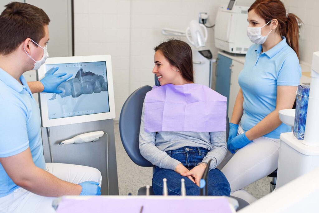
In addition, 3D printing technology has an advantage in printing smaller and more complex structures. It is recommended to combine the use of this new technology based on CBCT in the future, and it may even be used in other fields of oral and maxillofacial surgery, such as dental implant treatment, TMJ surgery, orthognathic surgery, and mandibular distraction osteogenesis. CBCT and 3D printing technology have broad application prospects in oral and maxillofacial reconstruction.



Leave a Reply